Experimental assessment of the effectiveness of electromagnetic therapy in purulent and burn wound healing process
Received:
Accepted:
Published:
Authors: Vishrabdha Chandrakant Margaj et al
Citation:
Chandrakant Margaj V et al, (2026) Experimental assessment of the effectiveness of electromagnetic therapy in purulent and burn wound healing process. Global Wound Care Journal 2 (2): 13-17.
Declaration of interest:
The authors have no conflicts of interest to disclose.
Corresponding author:
Margaj Vishrabdha Chandrakant Plastic Surgery and Laser Unit, Bedford Hospital South Wing, Bedford MK42 9DJ, UK. Email: Vishrabdha.Margaj@bedsft.nhs.uk
DOI: 10.63896/gwcj.2.2.13
Introduction: Wound infection is a significant healthcare problem, and electromagnetic therapy (EMT) is a promising tool to reduce bacterial load and promote wound healing.
Aim: This study aimed to provide an objective assessment of efficacy of EMT in healing of purulent and burn wounds in experimental animals.
Methods: Our prospective, non-randomised study enrolled 28 non-pedigree rabbits aged 2-3 months, divided into two groups. Wounds of the experimental group (n=14) were treated with local EMT using a TOR electromagnetic therapy device. The control group (n=14) was treated with 0.9% physiological saline solution. After 14 days of treatment, the wounds were assessed daily using planimetry and a bacteriological culture test. SPSS 17.0 was used to analyse the data.
Results: After 5 days of treatment, the experimental group was found to have a statistically significantly higher healing rate (p<0.05) as compared to the control group. At the same time, a positive correlation was noted between the speed of wound healing and the extent of the elimination of Gram-negative flora.
Conclusion: EMT has a broad-spectrum bactericidal effect on animals with purulent wounds and burns. It accelerates wound healing without any serious adverse effects.
Purulent wounds are a significant healthcare issue with a negative socio-economic impact. Approximately 35-45% of surgical patients have purulent wounds, often with polymicrobial flora (Peel et al, 2019). The ideal treatment method for such wounds needs to overcome resistance to antibiotics and stimulate the patient’s own regenerative resources.
Over recent years, electromagnetic therapy (EMT) has emerged as a promising tool for infected wounds because it achieves both goals (Pouriran et al, 2016 Bagheri et al, 2018; Kouhkheil et al, 2018; Dastgeyb et al, 2020; Bunkin et al, 2022).
The mechanism of action of EMT in healing purulent wounds is multifaceted. Electromagnetic fields improve microcirculation, ensure effective tissue oxygenation, and increase the bioavailability of antibiotics. It is anti-inflammatory and it stimulates fibroblasts, the cells responsible for collagen synthesis (Iryanov and Kiryanov, 2015; Yuan et al, 2015; Okhunov et al, 2018).
Clinical studies demonstrate promising results of the use of EMT when treating purulent wounds, with a notable wound contraction, reduction in duration of healing process and decrease in frequency of complications. EMT can be used both as monotherapy and in combination with other treatments, such as antibiotic therapy and surgical debridement (Bunkin et al, 2021; Ploskonos et al, 2022; Adams and Petruccione, 2024).
An important area of study is the influence of various electromagnetic field parameters, such as frequency, intensity and impulse form, on the speed and quality of regeneration and epithelisation (Betskii et al, 2000). The preliminary data indicate a significant stimulatory effect of low-frequency electromagnetic fields on wound constriction, but further research is necessary to confirm these results and optimise treatment parameters (Subbotina et al, 2006; Ziskin, 2013; Okhunov et al, 2018; Yuan et al, 2018).
Like any therapeutic treatment, EMT demonstrates a significant dose-dependent effect, which determines its effectiveness and safety. Dose dependence results from the complex interaction of electromagnetic fields with biological tissues. Notably, high doses may have a destructive effect on tissues due to excessive thermal effects, generation of free radicals and disruption of cellular structure (Lipatov et al, 2001; Ziskin, 2013).
Hence, the broad introduction of EMT into clinical practice requires further research, directed at optimising treatment parameters, developing new methods and assessing long-term effectiveness (Subbotina et al, 2006).
Aim
Our study aimed to perform an objective assessment of efficacy of EMT in healing of purulent and burn wounds in experimental animals.
Ethics committee approval
The conduct of the study was approved by the Local Ethics Committee of the Far Eastern State Agrarian University of the Russian Federation (Protocol No. 7 dated April 14, 2024).
The scientific studies were conducted in accordance with bioethical requirements, following the national general ethical principles governing experiments on animals and the relevant provisions of the European Convention for the Protection of Vertebrate Animals Used for Experimental and Other Scientific Purposes, dated March 18, 1986.
Methods
The prospective, non-randomised, open study was carried out from 01/06/2024 to 31/12/2024. It involved 28 non-pedigree rabbits, aged 2-3 months, weighing 1.5-2 kg, which were divided into two groups.
The experimental group (n=14) comprised animals with purulent and burn wounds that were treated with daily sessions of EMT. The control group (n=14) comprised animals with wounds treated by applying 0.9% physiological saline solution twice daily. Two subgroups were selected in each group depending on the etiopathogenesis of the infected wounds.
Subgroup A (n=7) was comprised of animals for whom purulent wounds were modelled by excising a skin flap measuring 25 cm² under local anaesthesia with infiltration of 0.25% novocaine solution. The skin flap was turned with the fur inside out and fixed with a plaster for 24 hours. As a result, signs of purulent inflammation formed on the second day. Subgroup B (n=7) comprised animals for which burn wounds were modelled by applying a red-hot brand to the skin with an area of 25cm2 for two seconds.
The “TOR” non-invasive electromagnetic therapy device was used to treat the wounds in experimental group. This device is registered as a medical device in the State Register of the Federal Healthcare Monitoring Service of the Russian Federation (Fatenkov et al, 2024). The electromagnetic therapy sessions were administered to the animals at a distance of 5m, daily at 12:00 pm, for 15 minutes.
The principle of the effect of the “TOR” device is based on a weak non-ionising nonthermal electromagnetic radiation, which is constantly generated by high voltage impulses from tungsten electrodes. The frequency of the impulses is in a range of 100-140 Hz, with each meander-like wave packet operating at a frequency of 25kHz. The power consumption during operation is 12 W with a maximum power of 150 W. The voltage on the tungsten does not exceed 8 kV. The electromagnetic radiation follows the standards of the National Healthcare Requirements of the Russian Federation.
Clinically, the body temperature of the animals was assessed every day with clinical and biochemical blood tests to analyse the markers of the systemic inflammatory response (blood leucocytes, neutrophil counts and C-reactive protein).
After the wound modelling, the daily measurement of the size of the wounds was carried out with the daily use of the planimetric method. The essence of this method was tracing the contour of the wound onto graph paper marked with a square grid. The number of squares that were within the boundaries of the wound were counted to calculate its area in cm2. This method allowed for the routine assessment of the dynamics of the size of the wound, including its depth and state of the wound bed.
A bacteriological test was carried out every day, by way of the standard method of taking a swab from the wound surface of the animal and placing it in a nutritious medium for the growth of bacteria, after which a microorganism culture was isolated and the further identification of the bacteria species, its concentration and sensitivity to antibiotics was performed.
The duration of the experiment was 14 days. SPSS 17.0 was used for the statistical processes of the data. The quantitative values are presented as the median and interquartile interval, Me [Q1; Q3]. The normality of the distribution of the qualitative values was carried out using the Shapiro–Wilk criteria. The statistical significance of the difference in the quantitative values between the three independent groups was assessed using the Craskell–Wallace criteria. To assess the correlation between the quantitative values, the Spearman rank correlation was used. The differences in categorical variables between independent groups were assessed using Pearson’s chi-squared test. Statistical significance was set at p=0.05.
Results
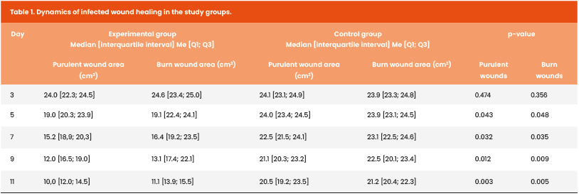
The rate of wound healing in the research group was assessed 5 days after the initiation of EMT. A statistically significant higher healing rate was observed in the experimental group [Table 1].
Before starting electromagnetic therapy, all animals displayed the presence of Gram-positive Staphylococci (Staphylococcus epidermidis, S aureus and S xylosus) at a maximum concentration of 104 CFU/ml. No statistically significant differences in their amounts were observed between animals with purulent and burn wounds during the study under electromagnetic therapy [Table 2].
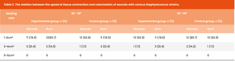
Cultures were negative in the experimental group on the 14th day, compared with 12 (85.7%) positive cultures in the control group. There was no correlation between a decrease in Staphylococcus bacteria and accelerated wound healing.
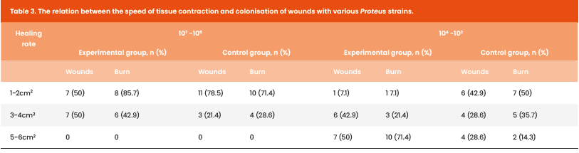
When analysing the Gram-negative flora, it was found that a decrease in the number of Proteus mirabilis and P vulgaris under treatment showed a statistically significant faster elimination and faster wound healing in the experimental group compared to the control group (p<0,05) [Table 3].
When assessing the degree of infection by Escherichia coli, Citrobacter freundii (coliform bacteria) and Klebsiella oxytoca, it was noted that they were equally widespread before the start of treatment. There was a notable progressive decrease in their presence in cultures, which was directly proportional to the treatment time [Table 4].
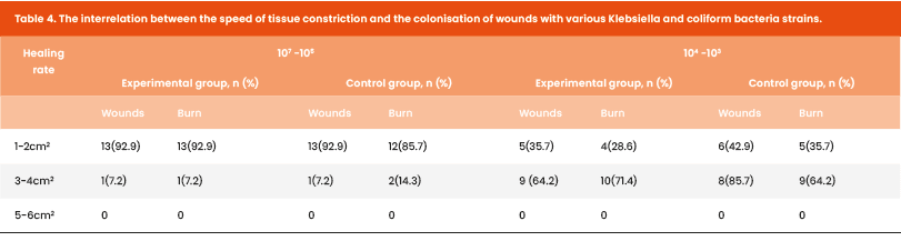
When analysing the speed of Acinetobacter species elimination in the experimental group, a rapid reduction in wound colonisation was observed after 5 days, with complete absence by the end of the experiment. It is important to note that with a decrease in Acinetobacter colonisation to 10³ CFU/ml, the rate of tissue regeneration increased by 50%. The elimination of this highly resistant microorganism proved extremely slow.
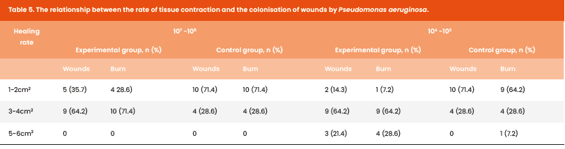
When analysing the speed of Pseudomonas aeruginosa elimination from the wound surface, it was noted that the treatment eliminated this microorganism from 10 (71.4%) animals within 10 days. A positive correlation was also observed between the wound-healing rate and the degree of elimination for this Gram-negative bacterium [Table 5].
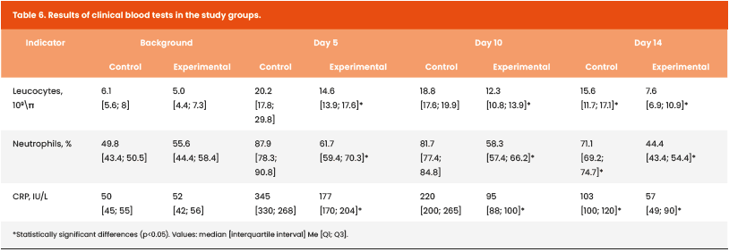
When analysing the clinical picture and laboratory results of the experimental animals, statistically significant differences in laboratory markers were observed between the study groups [Table 6]. Two peaks in leukocytosis and a decrease in C-reactive protein levels were noted in the experimental group. The second peak in CRP reduction on day 10 of the study was less pronounced and, in 11 (78.6%) cases, correlated with a decrease in P aeruginosa colonisation in the wounds.
Fever affected 11 (78.6%) animals in the experimental group and 12 (85.7%) in the control group during the first 5 days. Subsequently, all animals in both the experimental and control groups, regardless of wound aetiology, exhibited a subfebrile temperature up to 37.2°C for the remainder of the experiment.
Discussion
The speed of wound contraction is directly related to the harmonious progression of the acute phase of inflammation, exudation, proliferation and remodelling, and elimination of bacteria with the help of various treatment modalities.
The spectrum of the bactericidal effect of EMT is as broad as that of UV radiation, which today has become a widespread tool for bactericidal purposes. Prolonged, regular exposure to high doses of ultraviolet radiation may suppress immune cell activity, making the organism less resilient against other diseases, particularly skin cancers (Tsai and Chien, 2022; Park et al, 2023).
The EMT does not have these contraindications. The elimination of Gram-positive flora does not require high doses of radiation, although it does not qualitatively affect the rate of wound contraction in experimental animals. When analysing the bactericidal effect of EMT on Gram-negative flora, a decrease in their presence in cultures from wound and burn surfaces was directly proportional to treatment time. Hence, a direct relationship was observed between the rate of eradication of this Gram-negative flora and the rate of wound healing.
It is important to note that an electromagnetic field acts at the cellular level, disrupting bacterial metabolic processes and DNA structure. Unlike antibiotic therapy, EMT does not cause allergic reactions, does not damage healthy tissues, and bacteria do not develop resistance to it.
Therefore, electromagnetic therapy is a promising treatment for infected wounds and warrants further study. Its potential advantages, as well as its broad-spectrum bactericidal effect, safety and the possibility of combining it with other treatment methods, make it a promising field for the development of new approaches to combating surgical infections.
Conclusion
Electromagnetic treatment has a broad spectrum bactericidal effect on purulent wounds and burn wounds of animals. The mechanism of effectiveness of EMT is reduction of bacterial load and stimulation of tissue regeneration. During the experiment, no adverse effects were recorded.
Overall, EMT is a promising and innovative approach to the treatment of purulent wounds, which requires further research directed at optimising treatment parameters and an assessment of long-term effectiveness.