The eyebrow surprise: A rare giant lobular capillary haemangioma following trivial trauma in a child
Received:
Accepted:
Published:
Authors: P Umar Farooq Baba, Umesh K, Richam Kashyap, Jaswinder Kour, Adil Hafeez Wani
Citation:
Farooq Baba PU et al, (2026) The eyebrow surprise: A rare giant lobular capillary haemangioma following trivial trauma in a child. Global Wound Care Journal 2 (2): 39-42.
DOI
10.63896/gwcj.2.2.39
Background: Lobular capillary haemangioma, commonly known as pyogenic granuloma, is a benign but rapidly growing vascular tumour with a high tendency to bleed. Although frequently seen in the paediatric head and neck region, eyebrow involvement, especially of giant proportions, is extremely rare.
Case Presentation: We report a case of a 7-year-old boy who developed a 4 cm × 2.5 cm ulceroproliferative lesion over the lateral left eyebrow 2 weeks following trivial trauma. The initial laceration was sutured at a peripheral health centre, after which a rapidly enlarging mass developed over 2 weeks. Surgical excision with primary closure was performed. Histopathology confirmed lobular capillary haemangioma.
Conclusion: This case is unique due to its rare eyebrow location, large size, and post-traumatic onset, underscoring the need to consider lobular capillary haemangioma in the differential diagnosis of rapidly growing facial masses in children.
Lobular capillary haemangioma (LCH), formerly and misleadingly called pyogenic granuloma, is a benign capillary proliferation that typically arises in response to trauma, irritation or hormonal triggers. First described by Hartzell in 1904 (Hartzell, 1904), these lesions commonly occur in the skin and mucous membranes, especially the head and neck region in children and young adults, comprising up to 62% of reported cases (Patrice et al, 1991; Mills et al, 1980).
LCHs are notorious for their rapid growth and tendency to ulcerate and bleed, often causing diagnostic confusion with vascular neoplasms or even malignancies (Borden and Harrington, 2018). Lesions measuring over 2 cm are considered “giant” and are exceedingly uncommon in paediatric practice (Kheder et al, 2022). Eyebrow localisation is an extreme rarity, with only a single adult case reported involving this region in the literature (Bhavsar et al, 2024).
We present a case of a rapidly growing giant LCH over the lateral eyebrow of a child following minor trauma, adding to the limited spectrum of paediatric facial presentations of this entity.
Case report
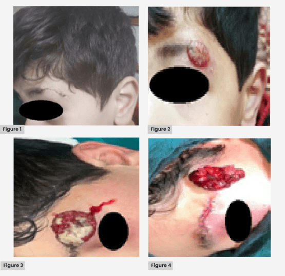
A 7-year-old boy presented to our outpatient department with a 4 cm × 2.5 cm ulcerated, friable mass over the lateral aspect of his left eyebrow. The child had sustained a minor blunt injury while playing at school. The trauma resulted in a small laceration approximately 1.5 cm in length over the left eyebrow region. He was taken to a nearby healthcare facility, where the wound was cleaned and closed with two simple interrupted nylon sutures. No foreign body was noted at that time. In the immediate post-injury period, the sutured wound appeared healthy with minimal swelling and no signs of infection [Figure 1]. By the fourth to fifth day, a small reddish nodule began to develop along the suture margin. At the time of suture removal on day 7, a tiny granulation-like swelling was noted. Over the next few days, the lesion enlarged rapidly and started bleeding on minimal contact. By the end of the second week, it had evolved into a pedunculated, ulcerated and friable mass with recurrent bleeding episodes, prompting referral to our institute. On presentation, the lesion measured 4 × 2.5 cm and appeared erythematous, lobulated and highly vascular, bleeding with trivial manipulation [Figure 2].
Pain and symptom management
The child experienced mild intermittent discomfort. It was managed with oral paracetamol (10 mg/kg) on an as-needed basis. No opioid analgesics were required.
Psychological aspects
The sudden appearance of a large bleeding facial lesion caused considerable anxiety to both the child and his parents. Preoperative counselling and reassurance were provided by the treating team, and the family was offered psychological support services. The child responded well to reassurance and supportive care.
Investigations
To exclude an underlying vascular malformation, a local colour Doppler ultrasound was performed, which demonstrated a superficial, low-flow vascular lesion without feeder vessels. Screening for visceral angiomas was carried out with abdominal ultrasonography, which was normal.
On examination, the lesion was pedunculated, erythematous, lobulated, and highly vascular, with focal ulceration and serosanguinous crusting [Figure 3]. The surrounding skin was unremarkable. There was no regional lymphadenopathy. No other systemic or cutaneous findings were noted.
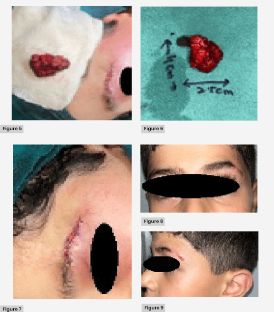
A clinical diagnosis of pyogenic granuloma (LCH) was made, with differentials including vascular malformation, foreign body granuloma and amelanotic melanoma. After an appropriate preoperative workup, the lesion was excised under general anaesthesia with meticulous haemostasis achieved throughout the procedure [Figures 4–6]. The surgical site was irrigated with sterile normal saline followed by 5% povidone-iodine solution to ensure optimal wound cleansing. The wound was closed primarily in layers, with meticulous haemostasis [Figures 4 and 5]. Postoperatively, a non-adherent sterile dressing was applied. Daily dressing changes were performed under aseptic conditions, with close monitoring for signs of infection or delayed healing. The wound healed uneventfully, and the postoperative course remained free of complications.
Histopathological Examination
The excised specimen was fixed in 10% buffered formalin and sent for routine histopathological examination. Paraffin-embedded tissue sections were prepared and stained using the standard Haematoxylin and Eosin (H&E) staining technique. No special stains, immunohistochemistry, transmission electron microscopy (TEM), or scanning electron microscopy (SEM) were performed.
Histopathological examination revealed lobular proliferation of capillary-sized vessels within a fibromyxoid stroma, with associated inflammatory infiltrate and surface ulceration – consistent with LCH. No cytologic atypia or mitotic activity was seen. However, histopathological photomicrographs were not available from the pathology department and, therefore, could not be included in this report.
The postoperative recovery was uneventful. At the 3-month follow-up, the child had a well-healed scar with no recurrence or residual deformity [Figures 7–9].
Discussion
LCH is a reactive vascular lesion resulting from angiogenic stimuli such as trauma, local irritation, or hormonal changes. Despite its name, it is neither pyogenic nor granulomatous in nature (Patrice et al, 1991). In children, the lesion is typically smaller than 1.5 cm and may be misinterpreted as a malignant or aggressive growth due to its alarming rate of expansion and propensity to bleed.
Trauma is a well-documented precipitating factor in paediatric LCH, reported in up to 30% of cases (Mills et al, 1980; Pagliai and Cohen, 2004). The most commonly affected sites include the lips, gingiva, cheeks and forehead. Eyebrow involvement is virtually unknown, with only one report describing a linear lesion in an adult male (Bhavsar et al, 2024).
Giant LCHs exceeding 2 cm are uncommon, and those ≥3 cm are exceedingly rare, particularly in paediatric populations. Thomas et al (2024) reported a case of giant pyogenic granuloma of the scalp in a 1-year-old child after trauma, underscoring the role of vascular hyperreactivity in this age group.
While topical timolol or imiquimod has shown promise in smaller lesions, surgical excision remains the mainstay for giant or ulcerated lesions, offering immediate resolution and histopathologic confirmation. Recurrence is rare after complete excision but may occur if the lesion is incompletely removed or subjected to partial treatments like curettage or cautery, which can have recurrence rates of up to 43% (Patrice et al, 1991; Lee et al, 2011).
Our case is notable for several reasons: it presents the rare occurrence of LCH in the eyebrow region, involves a giant lesion (>3 cm), and demonstrates a clear temporal relationship with minor trauma – factors that together make it a unique addition to the current literature.
Conclusion
LCH should be considered in the differential diagnosis of any rapidly growing, bleeding, or ulcerated lesion on the face, even in uncommon sites, such as the eyebrow. In children, minor trauma may suffice to trigger exuberant vascular proliferation, leading to diagnostic confusion. Complete excision with histopathologic confirmation ensures both therapeutic success and diagnostic certainty, especially in lesions of cosmetic or functional concern.
Patient consent
The authors certify that they have obtained all appropriate patient consent forms. In the form, the legal guardian has given consent for clinical information and images to be reported in the journal. The guardian understands that names and initials will not be published, and efforts will be made to conceal identity.
Ethical statement
The authors certify that they have obtained appropriate consent from the patient’s guardian. The patient’s guardian has given consent for images and other clinical information to be reported in the journal. The guardian understands that names and initials will not be published and due efforts will be made to conceal identity, but anonymity cannot be guaranteed. The study was conducted in accordance with the Declaration of Helsinki.